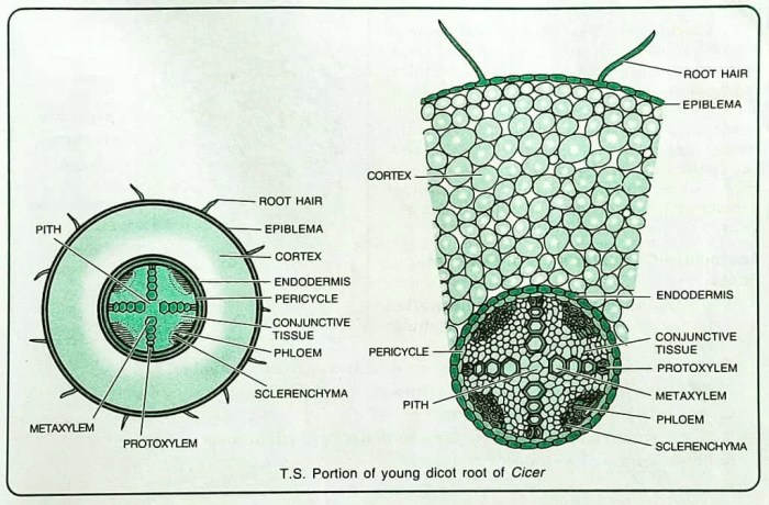
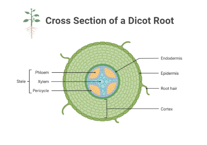
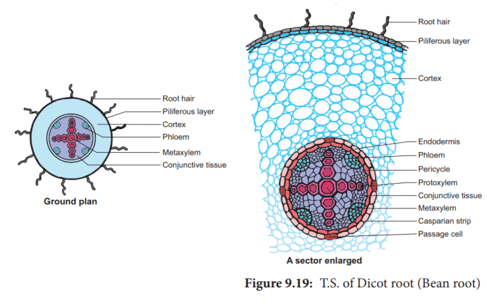
Cross section of a dicot root labeled – The cross section of a dicot root reveals a fascinating array of tissues and structures, each playing a vital role in the root’s function. From the protective epidermis to the nutrient-transporting vascular cylinder, this intricate anatomy provides a glimpse into the remarkable complexity of plant life.
The dicot root, a key component of the plant’s root system, serves multiple functions. It anchors the plant in the soil, absorbs water and nutrients, and stores food reserves. Understanding the cross section of a dicot root is essential for comprehending the root’s physiology and its significance in plant growth and development.
Introduction: Cross Section Of A Dicot Root Labeled

Studying the cross section of a dicot root provides valuable insights into the internal structure and organization of plant roots. Dicot roots, found in flowering plants, exhibit a distinct arrangement of tissues and structures that facilitate their various functions, including water and nutrient absorption, anchorage, and storage.
The cross section of a dicot root reveals several key tissues, each with specialized roles:
Epidermis
The epidermis is the outermost layer of the root and consists of a single layer of closely packed cells. It protects the root from the external environment and prevents water loss. The epidermal cells may have root hairs, which increase the surface area for water and nutrient absorption.
Cortex
The cortex lies beneath the epidermis and is composed of parenchyma cells, which are thin-walled and loosely arranged. The cortex stores food reserves and provides support to the root. It may also contain specialized cells, such as sclerenchyma cells, for added strength.
Endodermis
The endodermis is a single layer of cells that surrounds the vascular cylinder. It regulates the movement of water and nutrients into the vascular tissues. The endodermal cells have Casparian strips, which are waterproof barriers that prevent the passive movement of substances.
Pericycle, Cross section of a dicot root labeled
The pericycle is a layer of cells located outside the endodermis. It gives rise to lateral roots, which branch out from the main root and increase the root system’s surface area for water and nutrient absorption.
Vascular Cylinder
The vascular cylinder is the central region of the root and contains the vascular tissues: xylem and phloem. Xylem vessels transport water and minerals upward, while phloem tubes transport sugars and other nutrients downward.
Pith
The pith is the central core of the root and consists of parenchyma cells. It stores food reserves, such as starch, and provides support to the root.
Questions Often Asked
What is the significance of studying the cross section of a dicot root?
Studying the cross section of a dicot root provides insights into the root’s internal structure, tissue organization, and the functions of different tissues. It helps researchers understand how the root absorbs water and nutrients, transports them throughout the plant, and stores food reserves.
What are the main tissues found in the cross section of a dicot root?
The main tissues found in the cross section of a dicot root include the epidermis, cortex, endodermis, pericycle, vascular cylinder, and pith. Each tissue has a distinct structure and function, contributing to the overall functioning of the root.
How does the endodermis regulate the movement of water and nutrients?
The endodermis is a specialized layer of cells that surrounds the vascular cylinder. It contains a waxy substance called the Casparian strip, which restricts the movement of water and nutrients into the vascular cylinder. This regulation ensures that water and nutrients are transported through the selectively permeable cell membranes of the endodermal cells.

